Single Service
Open MRI
Service Information
Open MRI
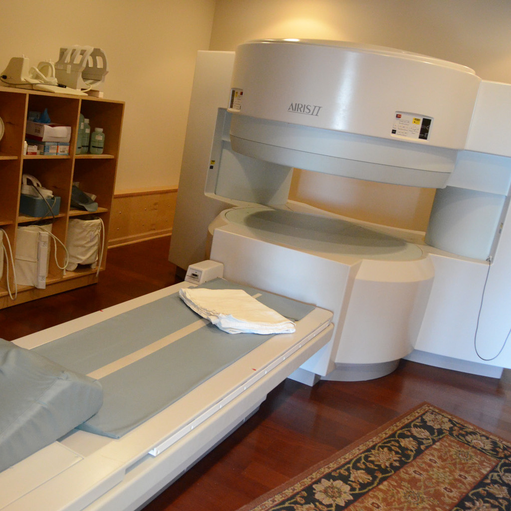
Our Open MRI captures high-quality images. Our technicians who operate the scanner are certified in MRI and will stay in constant communication with you throughout your scan.
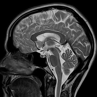
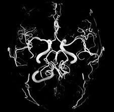
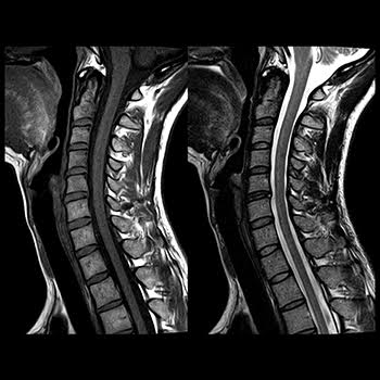
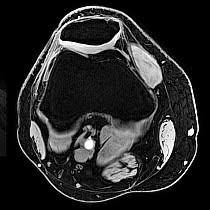
An MRI of the soft-tissue structures of the body—such as the heart, liver and many other organs - are more likely in some instances to identify and accurately characterize diseases than other imaging methods. This detail makes MRI an invaluable tool in early diagnosis and evaluation of many focal lesions and tumors.
Some of Your Questions
- What is MRI?
- How should I prepare for the procedure?
- How is the procedure performed?
- What will I experience during and after the procedure?
- Who interprets the results and how do I get them?
- What is MRI?
-
Magnetic resonance imaging (MRI) is a noninvasive medical test that helps physicians diagnose and treat medical conditions.
MRI uses a powerful magnetic field, radio frequency pulses and a computer to produce detailed pictures of organs, soft tissues, bone and virtually all other internal body structures. The images can then be examined on a computer monitor, transmitted electronically, printed or copied to a CD. MRI does not use ionizing radiation (x-rays).
Detailed MR images allow physicians to evaluate various parts of the body and determine the presence of certain diseases. - How should I prepare for the procedure?
-
You may be asked to wear a gown during the exam or you may be allowed to wear your own clothing if it is loose-fitting and has no metal fasteners.
Guidelines about eating and drinking before an MRI exam vary with the specific exam and also with the facility. Unless you are told otherwise, you may follow your regular daily routine and take food and medications as usual.
Some MRI examinations may require the patient to receive an injection of contrast material into the bloodstream. The radiologist or technologist may ask if you have allergies of any kind, such as allergy to iodine or x-ray contrast material, drugs, food, the environment, or asthma. The contrast material most commonly used for an MRI exam is called gadolinium. Because gadolinium does not contain iodine, it can be used safely in patients with contrast allergies.
The radiologist should also know if you have any serious health problems, or if you have recently had surgery. Some conditions, such as severe kidney disease may prevent you from being given contrast material for an MRI. If there is a history of kidney disease, it may be necessary to perform a blood test to determine whether the kidneys are functioning adequately.
Women should always inform their physician or technologist if there is any possibility that they are pregnant. MRI has been used for scanning patients since the 1980s with no reports of any ill effects on pregnant women or their babies. However, because the baby will be in a strong magnetic field, pregnant women should not have this exam unless the potential benefit from the MRI exam is assumed to outweigh the potential risks. Pregnant women should not receive injections of contrast material.
If you have claustrophobia(fear of enclosed spaces) or anxiety, you may want to ask your physician for a prescription for a mild sedative prior to the scheduled examination.
Jewelry and other accessories should be left at home if possible, or removed prior to the MRI scan. Because they can interfere with the magnetic field of the MRI unit, metal and electronic objects are not allowed in the exam room. These items include:
- jewelry, watches, credit cards and hearing aids, all of which can be damaged.
- pins, hairpins, metal zippers and similar metallic items, which can distort MRI images.
- removable dental work.
- pens, pocket knives and eyeglasses.
- body piercings.
In most cases, an MRI exam is safe for patients with metal implants, except for a few types. People with the following implants cannot be scanned and should not enter the MRI scanning area unless explicitly instructed to do so by a radiologist or technologist who is aware of the presence of any of the following:
- internal (implanted) defibrillator or pacemaker
- cochlear (ear) implant
- some types of clips used on brain aneurysms
- some types of metal coils placed within blood vessels
You should tell the technologist if you have medical or electronic devices in your body, because they may interfere with the exam or potentially pose a risk, depending on their nature and the strength of the MRI magnet. Some implanted devices require a short period of time after placement (usually six weeks) before being safe for MRI examinations. Examples include but are not limited to:
- artificial heart valves
- implanted drug infusion ports
- implanted electronic device, including a cardiac pacemaker
- artificial limbs or metallic joint prostheses
- implanted nerve stimulators
- metal pins, screws, plates, stents or surgical staples
In general, metal objects used in orthopedic surgery pose no risk during MRI. However, a recently placed artificial joint may require the use of another imaging procedure. If there is any question of their presence, an x-ray may be taken to detect and identify any metal objects.
Patients who might have metal objects in certain parts of their bodies may also require an x-ray prior to an MRI. You should notify the technologist or radiologist of any shrapnel, bullets, or other pieces of metal which may be present in your body due to accidents. Foreign bodies near the eyes are particularly important. Dyes used in tattoos may contain iron and could heat up during MRI, but this is rarely a problem. Tooth fillings and braces usually are not affected by the magnetic field, but they may distort images of the facial area or brain, so the radiologist should be aware of them.
Infants and young children usually require sedation or anesthesia to complete an MRI exam without moving. Whether a child requires sedation will depend on the child’s age and the type of exam being performed. Moderate and conscious sedation can be provided at most facilities. A physician or nurse specializing in the administration of sedation or anesthesia to children will be available during the exam to ensure your child's safety. You will be given special instructions how to prepare your child for the sedation or anesthesia. - How is the procedure performed?
-
MRI examinations may be performed on outpatients or inpatients.
You will be positioned on the moveable examination table. Straps and bolsters may be used to help you stay still and maintain the correct position during imaging.
photo of MRI procedure Devices that contain coils capable of sending and receiving radio waves may be placed around or adjacent to the area of the body being studied.
If a contrast material will be used in the MRI exam, a nurse or technologist will insert an intravenous (IV) catheter, also known as an IV line, into a vein in your hand or arm. A saline solution may be used. The solution will drip through the IV to prevent blockage of the IV catheter until the contrast material is injected.
You will be moved into the magnet of the MRI unit and the radiologist and technologist will leave the room while the MRI examination is performed.
If a contrast material is used during the examination, it will be injected into the intravenous line (IV) after an initial series of scans. Additional series of images will be taken during or following the injection.
When the examination is completed, you may be asked to wait until the technologist or radiologist checks the images in case additional images are needed.
Your intravenous line will be removed.
MRI exams generally include multiple runs (sequences), some of which may last several minutes.
Depending on the type of exam and the equipment used, the entire exam is usually completed in 15 to 45 minutes. - What will I experience during and after the procedure?
-
Most MRI exams are painless. However, some patients find it uncomfortable to remain still during MR imaging. Others experience a sense of being closed-in (claustrophobia). Therefore, sedation can be arranged for those patients who anticipate anxiety, but fewer than one in 20 require it.
It is normal for the area of your body being imaged to feel slightly warm, but if it bothers you, notify the radiologist or technologist. It is important that you remain perfectly still while the images are being recorded, which is typically only a few seconds to a few minutes at a time. You will know when images are being recorded because you will hear tapping or thumping sounds when the coils that generate the radiofrequency pulses are activated. You will be able to relax between imaging sequences, but will be asked to maintain your position without movement as much as possible.
You will usually be alone in the exam room during the MRI procedure. However, the technologist will be able to see, hear and speak with you at all times using a two-way intercom. Many MRI centers allow a friend or parent to stay in the room as long as they are also screened for safety in the magnetic environment.
You may be offered or you may request earplugs to reduce the noise of the MRI scanner, which produces loud thumping and humming noises during imaging. Children will be given appropriately sized earplugs or headphones during the exam. MRI scanners are air-conditioned and well-lit. Some scanners have music to help you pass the time.
In some cases, intravenous injection of contrast material may be performed. When the contrast material is injected in this fashion, it is normal to feel coolness and a flushing sensation for a minute or two. The intravenous needle may cause you some discomfort when it is inserted and once it is removed, you may experience some bruising. There is also a very small chance of irritation of your skin at the site of the IV tube insertion. Some patients may sense a temporary metallic taste in their mouth after the contrast injection.
If you have not been sedated, no recovery period is necessary. You may resume your usual activities and normal diet immediately after the exam. A few patients experience side effects from the contrast material, including nausea and local pain. Very rarely, patients are allergic to the contrast material and experience hives, itchy eyes or other reactions. If you experience allergic symptoms, notify the technologist. A radiologist or other physician will be available for immediate assistance. - Who interprets the results and how do I get them?
- A radiologist, a physician specifically trained to supervise and interpret radiology examinations, will analyze the images and send a signed report to your primary care or referring physician, who will share the results with you.
Follow-up examinations may be necessary, and your doctor will explain the reason why another exam is needed. Sometimes a follow-up exam is done because a suspicious or questionable finding needs clarification with additional views or a special imaging technique. A follow-up examination may be necessary so that any change in a known abnormality can be monitored over time. Follow-up examinations are sometimes the best way to see if treatment is working or if an abnormality is stable over time.
Phone: 708-301-4664
Fax: (708) 301-4641
Fax: (708) 301-4641